Kent Imaging: An Imaging Wonder in Wound Care and Reconstructive Surgery
Medical imaging technology in wound care and reconstructive surgery can make a world of difference in patient outcomes when the care providers are armed with timely, accurate, and actionable data. In essence, clinicians and their patients benefit from real-time insights into ‘what lies beneath’ wounded or questionable regions. Leading MedTech innovator, Kent Imaging, has been exemplary in driving clinical practices with innovations that help to reduce complications and improve results in wound and surgical care. Their flagship offering, SnapshotNIR, is a portable, easy-to-use, and non-invasive imaging device that eliminates the need for patient contact, leads, or injected dyes. This one-of-a-kind imaging device uses wavelengths of near-infrared (NIR) light to measure relative amounts of oxygenated and deoxygenated hemoglobin in tissue microcirculation in the wound area. It provides a near-instant tissue oxygen saturation map with a click of a button.
A Giant Leap in Imaging: SnapshotNIR
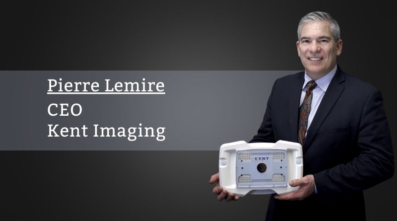
In the early 2000s, scientists at the National Research Council of Canada (NRC) created a prototype device to demonstrate how near-infrared light can effectively assess oxygen concentration in human tissue. Tissue oxygenation is one of the most important factors in wound healing, which cannot be measured by the naked eye. The NRC team demonstrated how multiple wavelengths of NIR light could reflect and measure the amount of oxygenated and deoxygenated hemoglobin in a patient’s microvasculature. Don Chapman, founder of Kent Imaging, knew the technology could improve people’s lives if made accessible. He secured the rights to the NRC’s laboratory model science and assembled a team at Kent Imaging to begin the development of a commercially viable NIR imaging unit. After many iterations, they went from a bulky stand model to a sleek handheld unit. Kent leveraged its progress by onboarding Pierre Lemire as CEO in 2016 to take the company to the next phase. Under Lemire’s guidance, Kent’s team continued to hone and improve—keeping Chapman’s original vision of an easily portable NIR tool as its objective.
“Tissue oxygenation is a key predictor of healing. Tissue health can provide valuable insight on whether a change of care plan is needed,” explains Pierre Lemire. “Snapshot uses NIR light to assess tissue oxygenation around the wound area. It shows whether the tissue is receiving the oxygen supply and if the oxygen is offloading into the tissue. These findings help confirm if the treatments for increasing tissue oxygenation positively impact the patient or if a new treatment plan is required.”
The company holds multiple patents in oxygen imaging technology and continues to provide advanced diagnostic imaging solutions to aid healthcare systems nationally and internationally. In 2017, Kent received FDA clearance and Health Canada certification for Snapshotto assess superficial tissue viability.Physicians have used Kent’s imaging device extensively to enable more efficient clinical decision-making and improve patient outcomes.
SnapshotNIR: a Revolution in Reconstructive Care
“Changes to a wound can be subtle and often not visible. Diagnostic-driven tools like Snapshotare valuable in understanding what is happening below the surface in the microvasculature network,” adds Lemire. In reconstructive surgery, the information obtained from the device can be extremely useful in the assessment of flap tissue viability. In under a minute, the images help physicians to identify whether the individual has adequate tissue oxygen saturation for flap survival, or if they may benefit from additional interventions. Surgeons can proceed confidently with their next steps.
SnapshotNIR’s greatest differentiator is the device’s ability to measure and report on the oxygen status of the microvascular network, where oxygen exchange occurs. It captures the oxyhemoglobin and deoxyhemoglobin status of the tissue imaged. Conventionally, imaging devices and products to assess tissue viability tend to be cumbersome, take considerable time to capture a study, and measure only macrovascular flow. The presence of blood, or even oxygenated blood, in the large vessels does not necessarily equate to oxygen in tissues. SnapshotNIR offers users rapid, actionable information that can impact decision-making and guide the treatment pathway.
Snapshot offers a truly different way to look at wound healing,” says Lemire.
“What makes our device stand out in the market is its capability to capture tissue oxygen saturation and hemoglobin data, as well as record wound area and linear measurements, all in one device,” explains Lemire. The ability to adjust the image contrast within the hemoglobin view allows device users to visualize the vasculature and the perfusion characteristics within the tissues of interest, providing clinicians and healthcare professionals additional metrics, above and beyond the tissue oxygenation view. When paired with the ease of Snapshot’s repetitive imaging abilities, users can quickly compare images pre- and post-intervention, at the bedside and without touching the patient, to assist in determining the potential of successful healing trajectories.
This tissue viability data captured on a regular basis can also be instrumental in confirming full wound healing. As an example, venous leg ulcers and diabetic foot ulcers have a high incidence of recurrence after healing. In the past, there has been no way to quantify the likelihood of reoccurrence, or if a wound is truly healed below the surface. Using Snapshot, wound specialists can continuously track and monitor the tissue healing to determine when the wound is healed on the surface and below as well. With this accurate and actionable data, rates of recurrence can be lowered, and clinicians can definitively decide on treatment adjustments.
“Snapshot offers a truly different way to look at wound healing,” says Lemire. “Our goal is for Snapshot imaging to become a standard in research for defining wound healing.”
Non-invasive and Cost-effective Imaging for Better Outcomes
Kent Imaging strives to deliver an innovative, cost-effective solution in an area where a rapid understanding of tissue viability and the ability to track and document wound healing progression can have a significant impact on healthcare costs, treatment decisions, and patient outcomes. The company is constantly working with established electronic health records (EHRs) to seamlessly integrate captured data from the device into the patient record. This integration ensures that critical information on the state of a wound is incorporated into the patient record at every visit, streamlining workflow while providing documented data to complete the patient chart.
Among its many use cases, Snapshot’s non-invasive tissue assessment capabilities have been leveraged to measure the effectiveness of the treatment pathway at the point of care and triage patients earlier in the care system. “If a treatment plan is ineffective or if new and significant insight is provided, clinicians can make timely changes to the treatment pathway and expedite referral to the appropriate specialty if required,” says Lemire. This was evident in the case of a patient with severe bilateral arterial insufficiency, and a previous right below-knee (BK) amputation. The patient developed a progression of her arterial disease with a breakdown of her BK amputation, resulting in a large ischemic wound. While the patient was being prepared for a right above-knee (AK) amputation, and in advance of the surgery, the Snapshot imaging captured and documented not only very severe ischemia in the BK wound, but it also indicated extremely poor oxygenation in the thigh. With this information, the physician was able to schedule an immediate vascular procedure (a right common femoral endarterectomy), as it was felt that the current tissue oxygenation in the thigh was inadequate to support an AK amputation. This additional vascular procedure would help establish adequate perfusion for the patient to support the healing of the AK amputation. This was a critical finding – had this issue not been recognized in advance and the amputation proceeded as originally scheduled, the AK amputation would likely have failed, leading to a large wound and potentially, a very poor outcome.
Trailblazers not Followers
At the core of Kent’s pioneering spirit is the drive to push boundaries to be at the forefront of innovation and technology. “We seek to improve the quality of life for individuals by bringing better medical solutions to all points of care. As a result, we are dedicated to overcoming challenges and pioneering a new way of visualizing disease,” says Lemire.
For the future, Kent Imaging aspires to be blazing the trail, not following it. “We focus on boldly experimenting and doing things that have not been done before. We love what we do, and we work hard to accomplish our goals. Passion is what fuels our drive,” says Lemire.